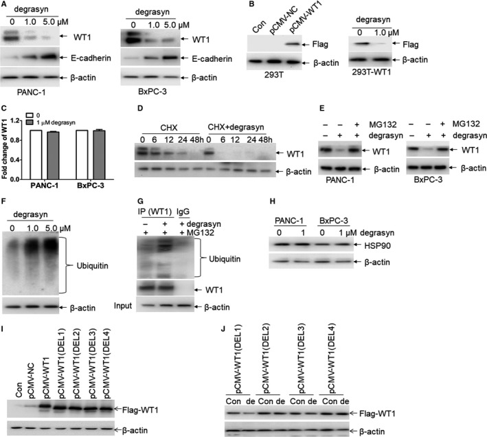
Figure 2.

Degrasyn induces the degradation of WT1 protein through enhancing ubiquitination. (A) The protein expressions of WT1 and E‐cadherin were measured in PANC‐1 and BxPC‐3 cells treated with or without 1 μM and 5 μM degrasyn for 24 hours. (B) Flag tag (Flag‐WT1) was detected in 293T cells transfected with pCMV‐NC or pCMV‐WT1 (Left blots). Flag tag was detected in 293T‐WT1 cells incubated with or without 1 μM degrasyn for 24 hours (Right blots). (C) The transcriptional expressions of WT1 were measured in PANC‐1 and BxPC‐3 cells treated with or without 1 μM degrasyn for 24 hours. (D) WT1 protein was detected in 10 μg/ml cycloheximide (CHX)‐treated PANC‐1 and BxPC‐3 cells, which were incubated with or without 1.0 μM degrasyn for the indicated times. (E) The protein expression of WT1 was measured in PANC‐1 and BxPC‐3 cells, which were treated with 1.0 μM degrasyn for 24 hours in the presence or absence of 5 μM MG132. (F) Ubiquitin was detected in PANC‐1 cells treated with 1.0 and 5.0 μM degrasyn for 4 hours. (G) PANC‐1 cells were treated with or without degrasyn, followed by the co‐immunoprecipitation (co‐IP) with anti‐WT1 antibody or normal mouse IgG. MG‐132 (5 μM) was added 4 hours before cell harvest. The Western blots were taken for ubiquitin and WT1 in co‐IP complex and input by anti‐β‐actin, respectively. (H) The protein expression of HSP90 was measured in PANC‐1 and BxPC‐3 cells, which were treated with or without degrasyn. (I) Flag tag (Flag‐WT1) was detected in 293T cells transfected with pCMV‐NC, wide‐type pCMV‐WT1 and four different pCMV‐WT1 deletions of zinc finger region. (J) 293T cells were transfected with four pCMV‐WT1 deletions for 24 hours and then treated with 1.0 μM WP1130 for 24 hours. Flag taq was measured by Western blot
