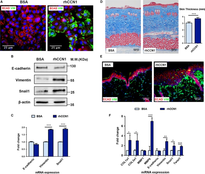
Figure 4.

rhCCN1 treatment induces partial EMT in rat keratinocytes and rat normal skin. A‐B, Primary keratinocytes were treated with rhCCN1 (0.2 µg/mL) for 48 h, and BSA (0.2 µg/mL) was used as a control. A, Immunostaining of the epithelial marker E‐cadherin (green) and mesenchymal marker Vimentin (red) (n = 3, scale bar: 25 µm). B, Western blotting of E‐cadherin, Vimentin and Snail1 (n = 3). C, Primary keratinocytes were treated with rhCCN1 (0.2 µg/mL) for 24 h. qRT‐PCR analysis of E‐cadherin, Vimentin and Snail1 (n = 3). D‐F, Rat normal skin was subcutaneously injected with rhCCN1 protein (2 µg/mL, 50 µL/d), and BSA (2 µg/mL, 50 µL/d) was injected as a control. D, Masson trichrome staining of the injected rat skin tissues in both groups (n = 5 per group, scale bar: 100 µm). E, Immunostaining of E‐cadherin (red) and Vimentin (green) in the injected rat skin tissues from both groups (n = 5 per group, scale bar: 50 µm). F, qRT‐PCR analysis of collagen production (COL1a1, COL3a1 and MMP1) and EMT markers (E‐cadherin, Vimentin, Snail1, Twist1 and MMP9) in the injected skin tissues from both groups (n = 5 per group). All values represent the means ± SD of triplicate determinations. *P < .05, **P < .01, ***P < .001
