
Figure 3.
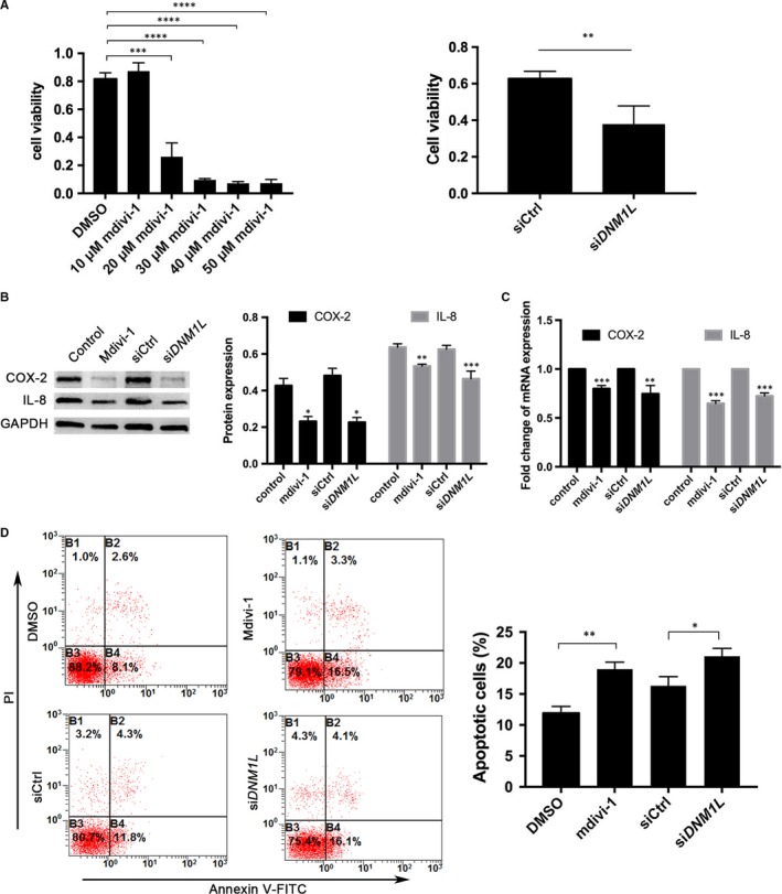
DNM1L deficiency in FLSs reduces their viability and production of pro‐inflammatory cytokines, and increases apoptosis. A, Cell viability was determined using the CCK‐8 assay. (B, C) Western blot and qRT‐PCR analyses of COX‐2 and IL‐8 expression in FLSs (mdivi‐1 concentration = 50 µmol/L). D, Representative flow cytometry data of apoptotic FLSs after staining with FITC‐Annexin V and PI, and quantitation of these data. Data are representative flow cytometry charts, images or expressed as mean ± SD of each group from three separate experiments. *P < .05; **P < .01; ***P < .001; ***P < .0001