
Figure 2.
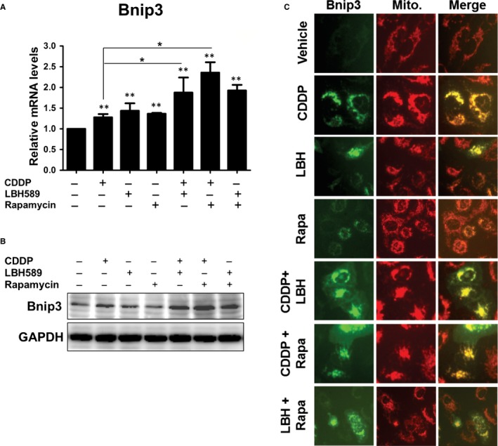
Increased levels and mitochondria localization of BNIP3 were identified upon treatment with anticancer drugs. A, B, A549 cells (2 × 10⁵) were treated with cisplatin (2 μg/mL), LBH589 (100 nmol/L), or rapamycin (100 nmol/L), or a combination of two drugs for 24 h (A) or 48 h (B). The mRNA or protein levels of BNIP3 were detected by qRT‐PCR (A) or Western blot (B), respectively. C, A549 cells were transfected with pEGFP‐BNIP3 plasmid. Sixteen hours later, cells were treated with anticancer drugs for 24 h. Fluorescent dye‐labelled mitochondria were detected using fluorescence microscopy upon treatment of 2 nmol/L of tetramethylrhodamine methyl ester perchlorate (TMRM) for 30 min. Results are representative of at least three independent experiments. **P < .01 compared with untreated cells