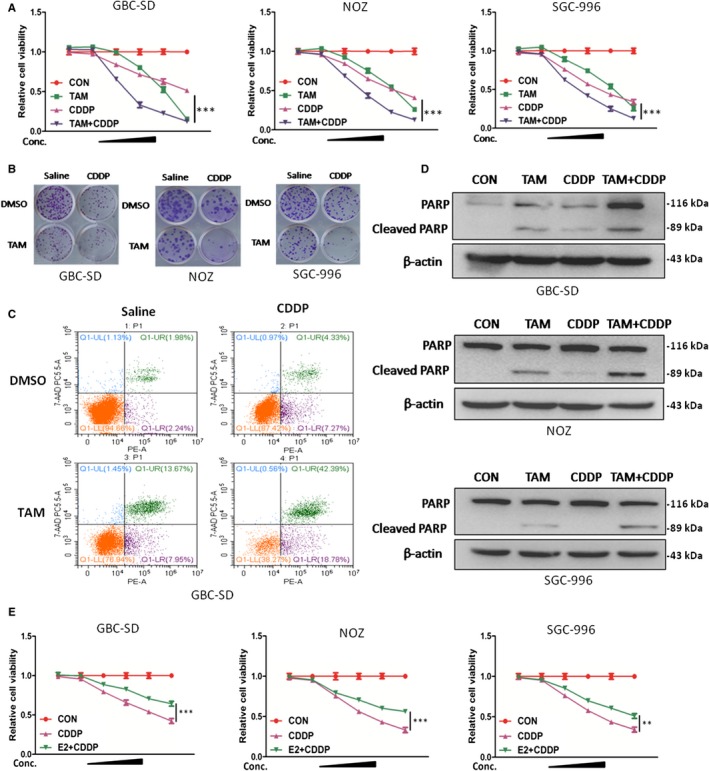
Figure 4.

TAM enhanced CDDP‐induced inhibition of cell viability in GBC cells. A, Cell viability in GBC‐SD,NOZ and SGC‐996 cells treated with TAM alone, CDDP alone, and TAM/CDDP combination for 48 h. TAM concentrations:0, 5, 7.5, 10, 12.5 and 15 μmol/L; CDDP concentrations: 0, 1, 2, 4, 8 and 12 μm. B, Representative images of colony in GBC cells. Cells were treated with TAM alone (5 μmol/L), CDDP alone (2 μmol/L) and TAM/CDDP combination for 24 h and incubated in the plate for 14 d. C, Apoptosis rate analysis using PE/7‐AAD flow cytometry in GBC‐SD cells treated with TAM alone (12.5 μmol/L), CDDP alone (4 μmol/L), and TAM/CDDP combination for 48 h. D, Protein levels of PARP and cleaved PARP in GBC cells treated with TAM alone (12.5 μmol/L), CDDP alone (4 μmol/L) and TAM/CDDP combination for 48 h. E, Cell viability in GBC‐SD,NOZ and SGC‐996 cells treated with E2 alone, CDDP alone, and E2/CDDP combination for 48 h. E2 concentrations: 0, 50, 100, 200, 400 and 800 nmol/L; CDDP concentrations: 0, 1, 2, 4, 8 and 12 μmol/L. TAM, tamoxifen; CDDP,cisplatin; E2, 17β‐estradiol,β‐actin was used as control in Western blot assay. All n = 3; bar, SEM
