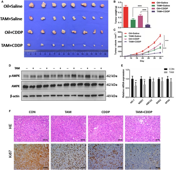
Figure 5.

TAM markedly sensitized the tumour xenografts to CDDP cytotoxicity in vivo. The tumour ‐bearing mice were injected intraperitoneally with dissolvents, TAM alone, CDDP alone and TAM/CDDP co‐administration. A, Photograph of transplanted tumours after the mice were exposed to treatments (n = 8/group). Oil and peanut oil. B, Tumour weight of four groups after the mice was exposed to treatments. C, Tumour growth curves of GBC‐SD cells after treatment in vivo. D, Western blot to analyse p‐AMPK and AMPK protein expression in transplanted tumours. E, Q‐PCR to analyse HO‐1, NQO1, ABCG2, SOD2 and GPX4 mRNA levels in transplanted tumours. F, H&E staining and Ki67 immunostaining in transplanted tumour tissues. Scale bars: 50 μm. TAM, tamoxifen; CDDP, cisplatin; Q‐PCR data were normalized by GAPDH, and β‐actin was used as control in Western blot assay. All bar, SEM, *P < .05; **P < .01; ***P < .001; Student's t test
