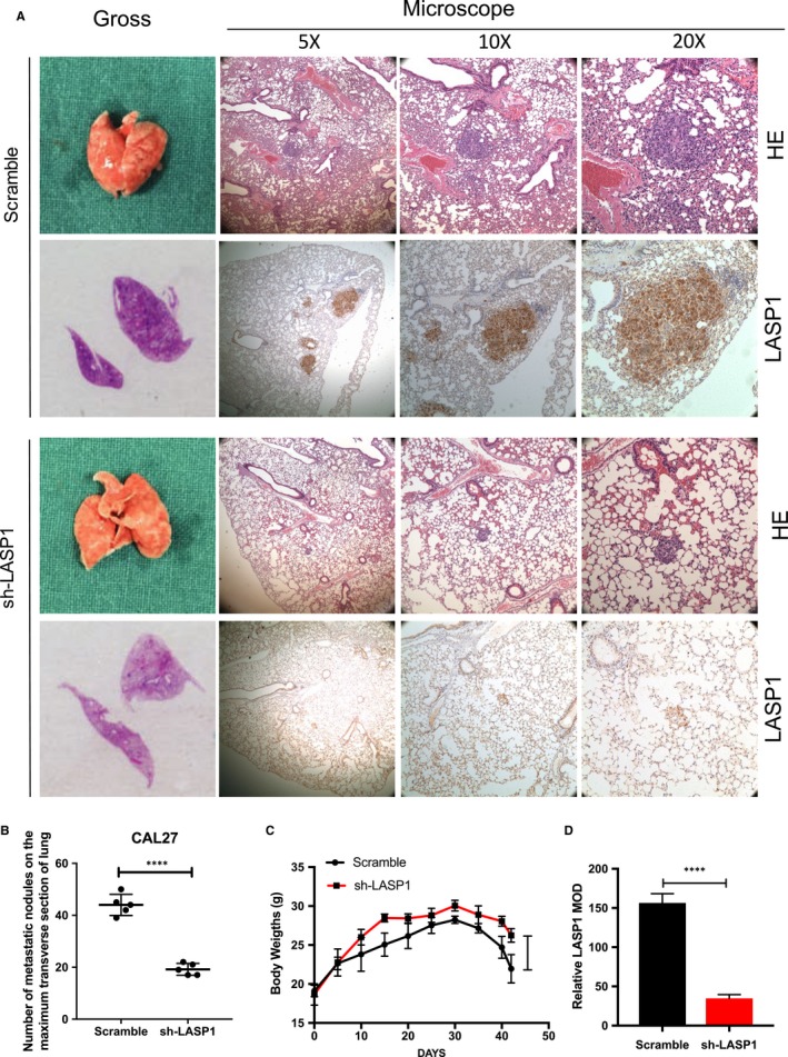
Figure 4.

LASP1 overexpression promotes HNSCC cell migration and invasion in vivo. A, The representative images of lung tissues of nude mice. After the HNSCC CAL27 cells (1 × 10⁶) were injected into the nude mice via the tail vein for 42 d, the lung tissues were fixed and examined grossly and then under the microscope (5×, 10×, 20×). H‐E staining was performed, and IHC staining was used to detect the expression of LASP1 in metastatic lesions. B, The quantity of metastases node in the largest transverse section of the lungs in the nude mice (n = 5 per group). ****P < .01 compared with the Scramble group. C, The body weight change of the nude mice (n = 5 per group) ****P < .01 in the interference group compared with the Scramble group. D, The relative number of LASP1 MOD between the interference group and the Scramble group was observed using ImageJ software. (n = 5 per group) ****P < .01
