Figure 6.

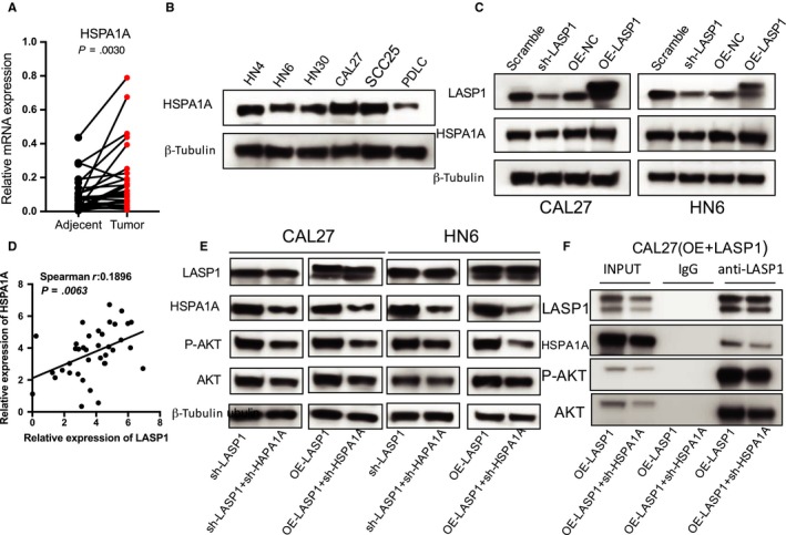
HSPA1A positively regulates LASP1‐pAKT directly binding. A, The real‐time PCR analysis of HSPA1A mRNA expression in 40 pairs of HNSCC tissue specimens and adjacent normal tissues (P = .0030). B, The Western blot analysis of HSPA1A levels in the HNSCC cell lines and PDLC cells. C, The Western blot analysis revealed that HSPA1A protein levels were significant altered in HNSCC cells (Scramble, sh‐LASP1, OE‐NC, OE‐LASP1). D, A significant positive correlation was observed between the LASP1 and HSPA1A expression levels in the HNSCC tissues (n = 40). E, Western blot analysis was performed to detect the HSPA1A interference efficiency and the influence of HSPA1A interference on the protein expression of P‐AKT and AKT. F, A Co‐IP analysis data were performed. The CAL27 (OE‐LASP1) cells were lysed and purified with an anti‐LASP1 affinity gel. The protein pellets were analysed by immunoblotting with anti‐LASP1, anti‐HSPA1A, anti‐P‐AKT and anti‐AKT. *** P < .05, **** P < .01
