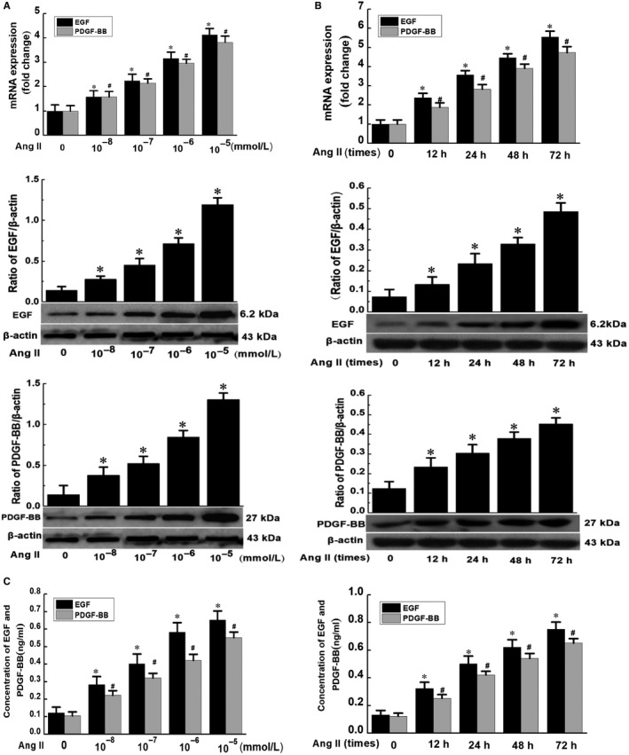
Figure 1.

Effect of Ang II on EGF and PDGF‐BB in phenotypic transformation of VSMCs. A, VSMCs were stimulated with different concentrations of Ang II for 48 h; B, VSMCs were stimulated with Ang II (10−5 mmol/L) for different durations. The total RNA was extracted, and the cDNA was obtained after reverse transcription. The expressions of EGF and PDGF‐BB were detected by RT‐qPCR. The total protein was extracted, and the expressions of EGF and PDGF‐BB at the protein level were analysed by Western blotting analysis. (Data were expressed as ± S, n = 3; * and # P < .05 vs the control group.) (C) After treatment of VSMCs with Ang II at different concentrations and durations, the cell culture supernatant was collected, and the secretion of EGF and PDGF‐BB was detected by ELISA. (Data were expressed as ±S, n = 3; * and # P < .05 vs the control group)
