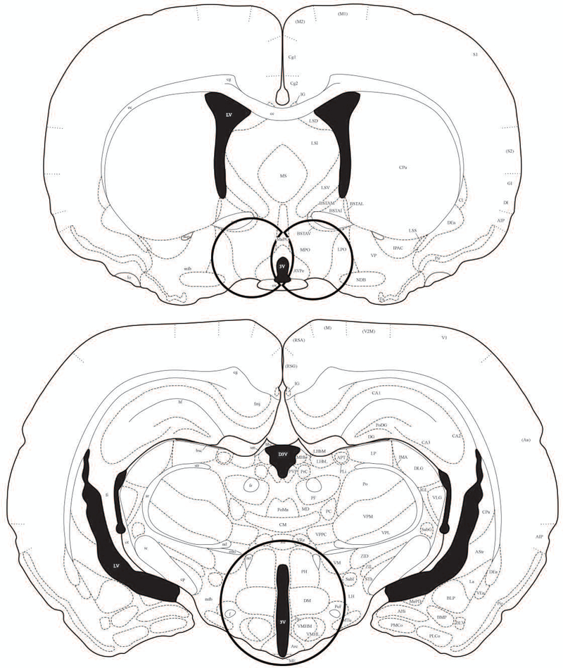
Figure 1.
An illustration of the location of samples punched for RT-PCR analysis. Brains were flash frozen and cut at 300μm and then transferred to RNAlater for one night. A 2mm biopsy punch was used to microdissect the mPOA and AVPV bilaterally (left), and a 3mm biopsy punch was used to microdissect the DMH and ARC in a single punch (right). Illustrations adapted and modified from the Stereotaxic Atlas of the Golden Hamster Brain by L.P Morin and R.I Wood (2000) (68).

