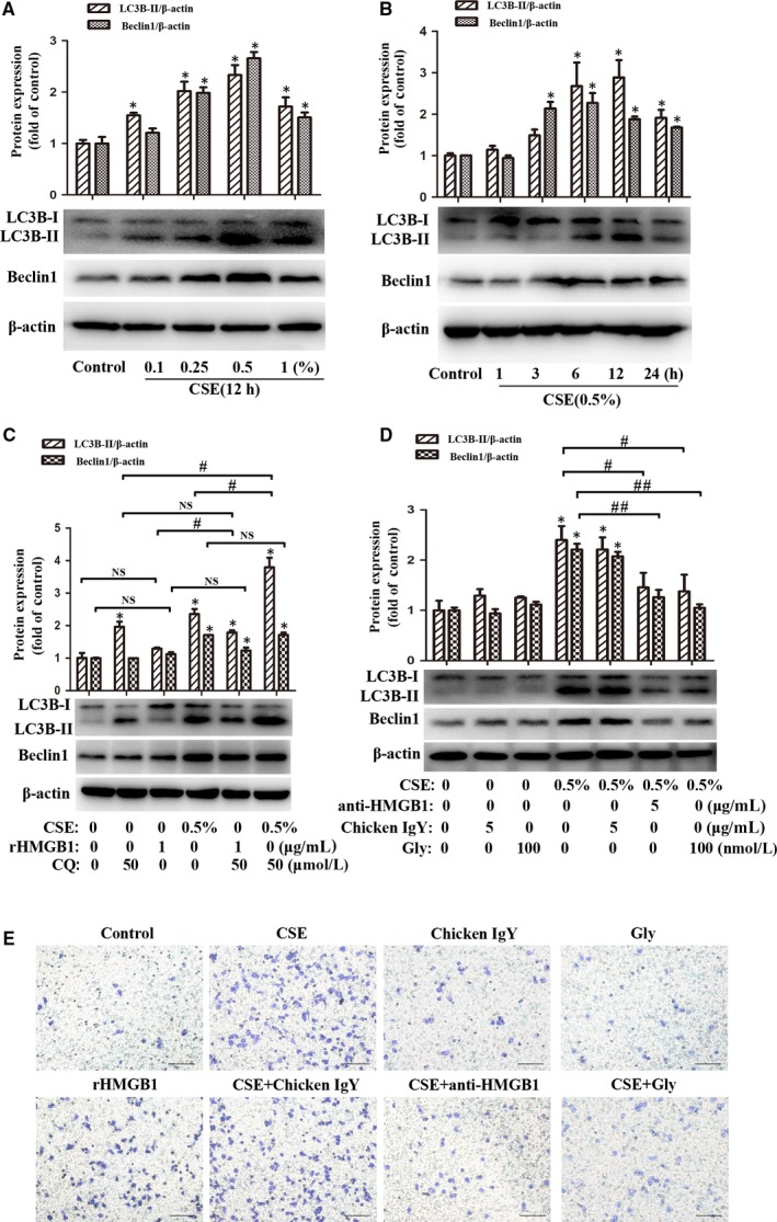
Figure 5.

Blockade of HMGB1 attenuated CSE‐induced autophagy and migration of lung macrophages. A, After MH‐S cells were treated with CSE (0, 0.1%, 0.25%, 0.5% or 1%) for 12 h, LC3B‐I/ II and Beclin1 were detected with Western blot. B, After MH‐S cells were treated with 0.5% CSE for 1, 3, 6, 12 or 24 h, LC3B‐I/ II and Beclin1 were detected with Western blot. C, After MH‐S cells were pre‐treated with CQ (50 μmol/L) for 2 h, CSE (0.5%) or rHMGB1 (1 μg/mL) were then added to stimulation for 12 h. D, MH‐S cells were pre‐treated with chicken IgY (5 μg/mL), anti‐HMGB1 (5 μg/mL) or Gly (100 nmol/L) for 90 min before 0.5% CSE incubated for 12 h. E, MH‐S cells were pre‐treated with chicken IgY (5 μg/mL), anti‐ HMGB1 (5 μg/mL) or Gly (100 nmol/L) for 90 min before 0.5% CSE or rHMGB1 (1 μg/mL) alone incubated for 24 h. Migration assay was assessed. Bar: 100 μm.*P < .05 vs Control group. # P < .05. ## P < .05. NS, no significant. Values are mean ± SEM of three independent experiments
