
Figure 5.
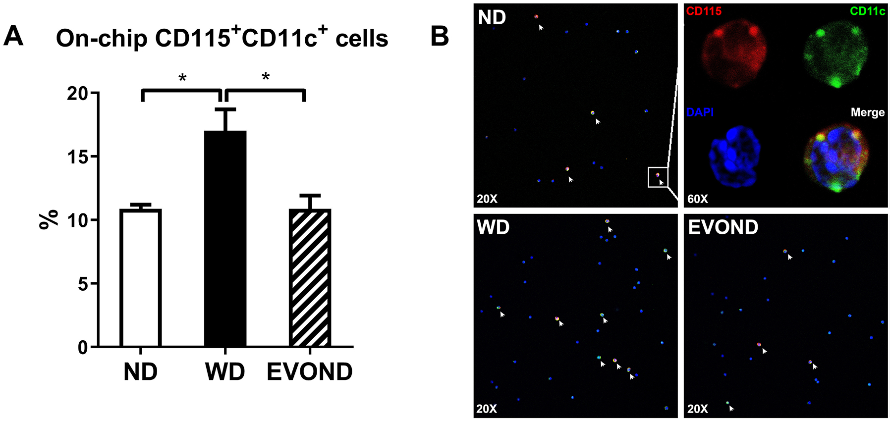
Reduced on-chip adhesion of foamy monocytes from mice on EVOND vs. WD. Circulating monocytes were stained with CD115 and CD11c and perfused through VCAM-1– and E-selectin–coated chip. The number of arrested CD11c+ monocytes were counted and normalized by total infused monocyte number. A, Frequency of CD11c+ monocytes arrested on chips in the total infused monocyte. Data are shown as mean±SEM, *p<0.05, n=4/group. B, Representative images showing arrested cells on chips under flow with FITC-anti-CD11c (Green), PE-anti-CD115 (Red), and DAPI (Blue) staining. See “Ex vivo micro–flow adhesion assay” under Materials and Methods for experimental procedures. Foamy monocytes marked with white arrows were identified by CD115 and CD11c double staining.