
Figure 6.
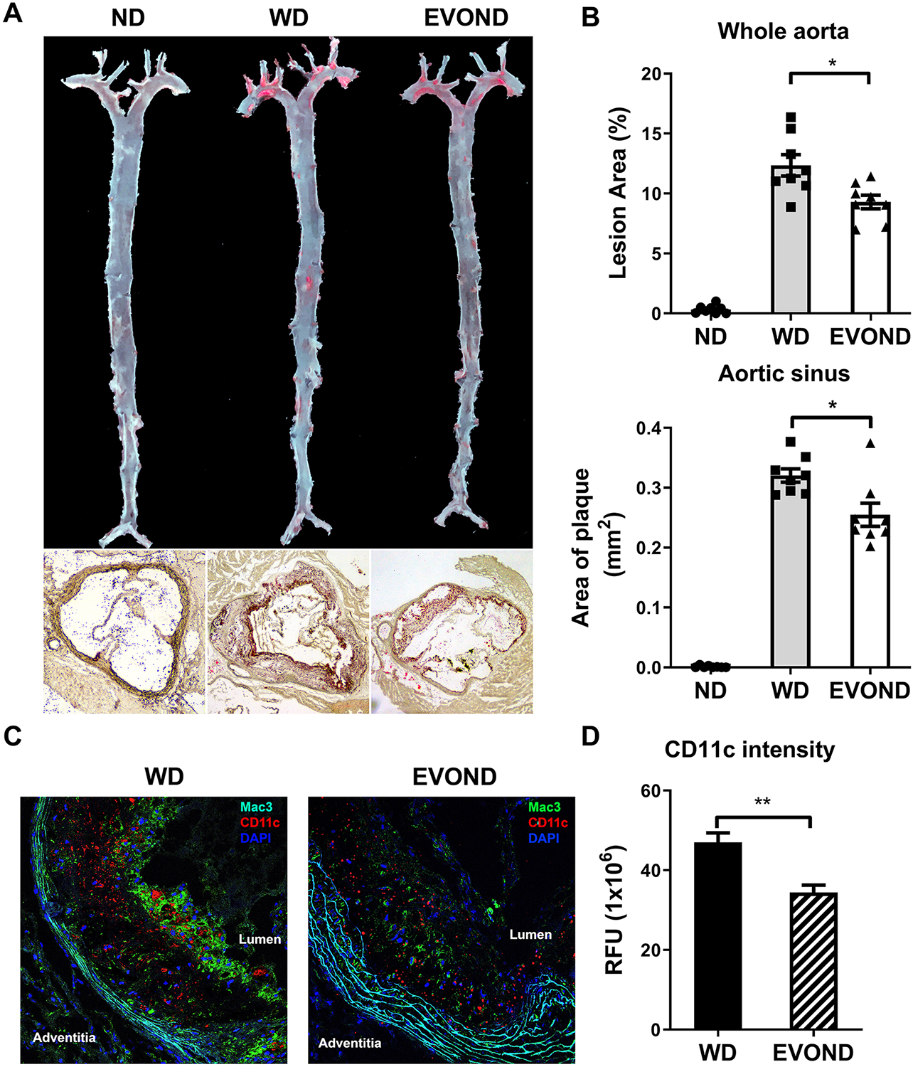
EVOND reduced atherosclerosis compared to WD in Ldlr–/– mice on diets for 3 months. A, Representative en face oil red O staining of whole aorta and aortic sinus (10x original magnification). B, Statistics of plaque area of whole aorta (upper panel) and aortic sinus (lower panel) (n=8/group). C, Representative immunofluorescence staining of Mac3 (green) and CD11c (red), with DAPI staining (blue) for nuclei, in aortic sinus lesions. D, CD11c fluorescent intensity (relative fluorescent unit [RFU]) in plaque of mice from WD and EVOND groups (n=6/group). Data are shown as mean±SEM. *p<0.05, **p<0.01.