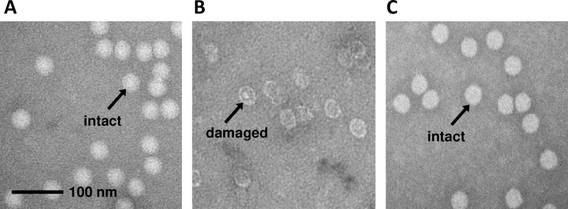
Fig 3. Ultrastructural analysis of irradiated poliovirus.
Electron micrographs are shown of PV1-S that had been A) not irradiated, B) irradiated to 45 kGy without MDP, or C) irradiated to 45 kGy in the presence of MDP. The images are representative of >700 particles assayed for each group. The length of the scale bar in Panel A is 100 nm.

