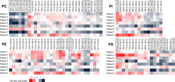
Fig 3. Tumor/non-tumor (T/N) ratios of specific PL species in patient-derived primary epithelial cells.
T/N ratio of chromatogram peak areas summarized according to MWs of lipid species (log transformation) in CRC patients (n = 8). Carbon and DB numbers are shown in parentheses. White color (zero value)—no difference between peak areas (= lipid amount) in tumor and non-tumor cells of the patient. Values lower than zero (red color)—lower amount of the respective PL species in tumor cells as compared with non-tumor cells. Values higher than zero (grey color)—higher amount of the respective PL species in tumor cells as compared with non-tumor cells. PL species with significant difference between tumor and non-tumor cells (the most discriminating) are highlighted in black frames (paired t-test).

