
Figure 3. Anti-proliferative efficacy of TAP NPs in breast cancer cells.
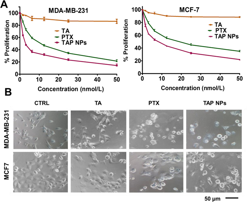
Cell proliferation measured by MTT assay. A) TAP NPs markedly decreased proliferation of breast cancer cells (MDA-MB-231 and MCF7) after treatment with 1–50 nmol/L PTX or TAP NPs (equivalent amount of drug) for 48 hours. Untreated cells were used as control. Effect of TAP NPs and PTX were assessed by MTT reagent and absorbance was recorded by a Microplate Reader (BioTeK Cytation 3, Winooski, VT, USA) at 570 nm. Data presented as mean ± standard error of the mean (n = 3). B) Bright field microscopy images after MTT analysis show significant changes in cell morphology after 48 hours treatment with TAP NPs and PTX. Imaging was done by an EVOS® FL Imaging System (20X magnification, scale = 200 μm).