
Figure 8. Molecular mechanism of TAP NPs in inducing apoptosis in breast cancer cells.
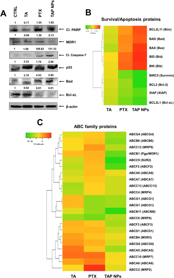
A) Western blot analysis of whole cell lysates of MDA-MB-231 that was treated with 10 nmol/L PTX or 10 nmol/L PTX equivalent TAP NPs and their respective controls for 48 hours and immunoblotted for Cleaved PARP, MDR1/ABCB1, Cleaved Caspase-7, p53, Bad, Bcl-xL and β-actin. The results were consistent in two independent sets of experiments. B) Heat map of differentially regulated gene expression of pro-apoptotic and anti-apoptotic signaling leading to apoptosis in MDA-MB-231 breast cancer cells after exposure to TAP NPs. C) Heat map representing gene expression of ABC family.