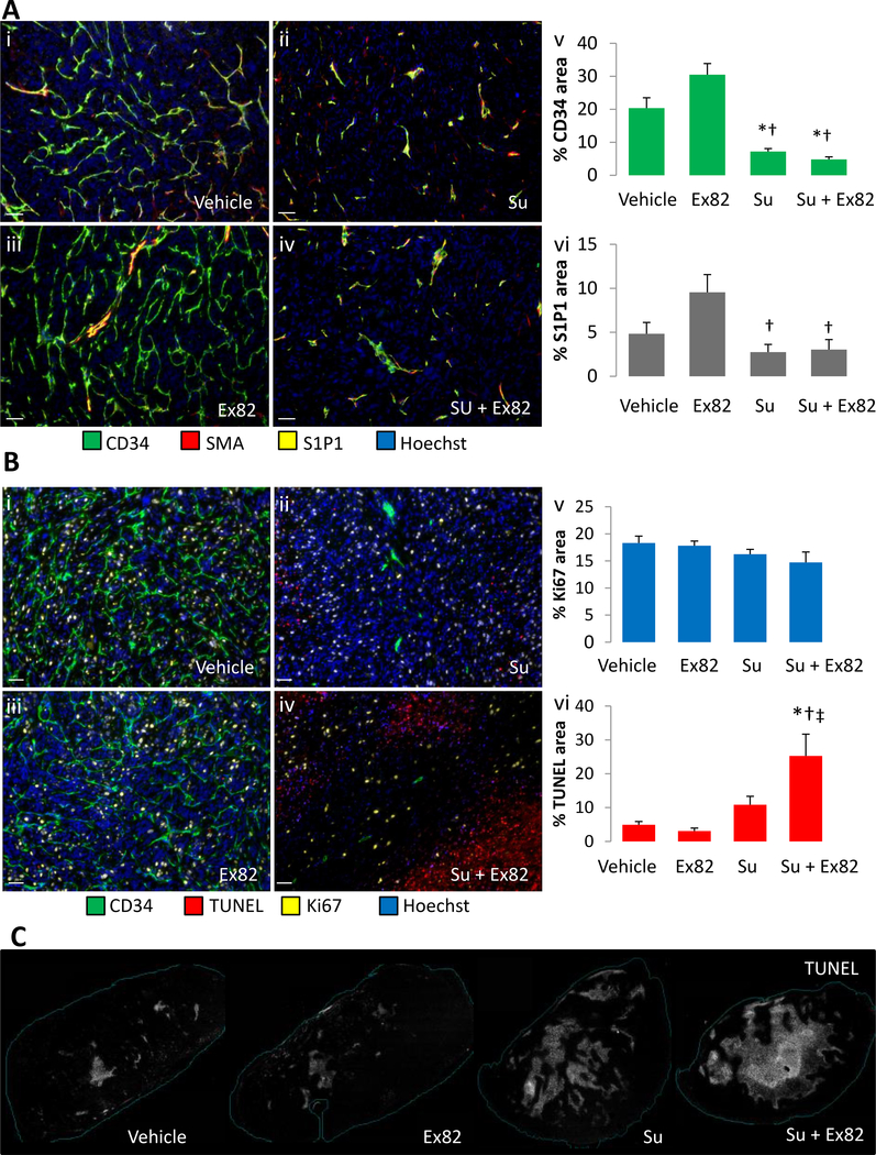
Figure 6. The combination of VEGFR and S1P1 inhibition induces tumor cell apoptosis.
Multiplexed panels to assay tumor angiogenesis are shown. The percent area of tumor vessels (labeled with CD34; green) increased with Ex82 (6Aiii and 6Av) and sunitinib (Su) significantly reduced the percent area of vessels (6Aii and 6v), but further reduction in tumor vessels was not detected with the combination of Ex82 and sunitinib (6Av). Pericyte staining was assessed by SMA (red), Hoechst staining is shown in blue, and S1P1 is shown in yellow. S1P1 expression tended to increase with Ex82 (6Aiii and 6vi) and was significantly less with sunitinib and the combination treatment. Examination of the effects of treatment on tumor cells showed that sunitinib treatment tended to increase apoptosis (TUNEL stain shown in red) and the combination treatment significantly increased apoptosis more than the vehicle or either of the single agents (6Biv and 6vi). CD34 staining is shown in green and Ki67 is shown in yellow. Hoechst staining is shown in blue. Whole tumor cross-sections are shown in 6C stained for TUNEL (gray). Bars represent mean +/− SEM. * = p<0.05 vs. vehicle, † = p<0.05 vs. Ex82, ‡ = p<0.05 vs. Su.

