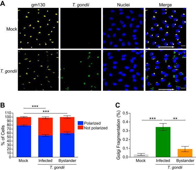
FIG 5.
Effect of T. gondii infection on HUVEC planar cell polarity. HUVEC were cultured to confluence on ibiTreat 0.4-luer ibidi microchannels under 5.5-dyne/cm2 shear stress for 3 days and either mock infected with fresh media or infected with T. gondii. At 18 hpi, the samples were fixed, permeabilized, and stained with anti-gm130 to detect the Golgi complex. (A) Fluorescence microscopy of gm130, GFP-expressing T. gondii, and nuclei at 5.5-dyne/cm2 shear stress in mock-infected and T. gondii-infected cells at 18 hpi is shown. The arrows in the merged images indicate the direction of flow. Bars = 20 μm. (B) Planar cell polarity of mock-infected and T. gondii-infected HUVEC at 18 hpi is shown. Combined data from three independent experiments are presented as the means ± SEM. One-way ANOVA with a Tukey posttest correction was performed (***, P < 0.001). (C) The percentage of cells exhibiting Golgi fragmentation was quantified in the mock-infected and T. gondii-infected cultures at 18 hpi. Directly infected cells and bystander cells in the infected cultures are shown. Data are presented as means ± SEM from three independent experiments. One-way ANOVA with a Tukey posttest correction was performed (***, P < 0.001; **, P < 0.01).

