. 2019 Dec 6;8:e46842. doi: 10.7554/eLife.46842

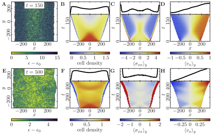
Figure 5. Expansion of a confluent epithelial cell sheet after removal of boundaries positioned at x=±175 for two different parameter settings.

(Stiffness parameters κP=0.12κA=0.18; average polarization field ϵ0=35; signaling radius R=2; cytoskeletal update rate μ=0.1; cell-cell adhesion B=12; cell-cell dissipation ΔB=0; cell-substrate dissipation D=0; cell-substrate adhesion penalty φ=0100 independent simulations for each set of parameters). (A–D) Tissue expansion for a migration-dominated setup without explicit cell growth and mitosis. (3300-cell system; maximum cell polarity Δϵ=30). (E–H) Tissue expansion at low density and cell polarizability for a cell sheet comprised of dividing cells. (Initially a 2500-cell system; maximum cell polarity Δϵ=10; growth time Tg=180; division time Td=20; size threshold for cell growth AT=1Aref, where Aref is the size of a solitary cell in equilibrium). (A, E) Snapshots of the polarization field ϵ; cf. Figure 5—video 1 and Figure 5—video 2. (B, F) Kymographs showing the cell density averaged over the y-direction and (top) final snapshots of the cell density profiles. (C, G) Kymographs showing the component σxx of the stress tensor averaged over the y-direction and (top) final snapshots of the stress profiles. (D, H) Kymographs showing the component vx of the cell velocities averaged over the y-direction and (top) final snapshot of the velocity profiles.

Figure 5.

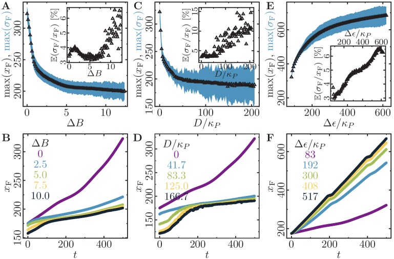
Figure 5—figure supplement 1. Monolayer expansion depends on dissipation and cell polarizability.

Figure 5—figure supplement 1.

Cell monolayer expansion depends on the cell-cell dissipation, cell-substrate dissipation, and maximum cell polarity. (Initially a 2500-cell system; stiffness parameters κP=0.12, κA=0.18; average polarization field ϵ0=35; maximum cell polarity Δϵ=10; signalling radius R=2; cytoskeletal update rate μ=0.1; cell-cell adhesion B=12; cell-substrate adhesion penalty φ=0; growth time Tg=180; division time Td=20; size threshold for cell growth AT=1Aref, where Aref is the size of a solitary cell in equilibrium; 100 independent simulations for each set of parameters). (A,B) Cell monolayer expansion depends on the cell-cell dissipation ΔB (maximum cell polarity Δϵ=10; cell-substrate dissipation D=0). (A) Maximal monolayer extension and roughness (we exclude an initial time interval of 200 MCS because it takes at least that long for first daughter cells to appear). Inset: Relative roughness of the spreading monolayer relative to its size. (B) Time traces for selected values of ΔB. (C,D) Cell monolayer expansion depends on the cell-substrate dissipation D (maximum cell polarity Δϵ=10; cell-cell dissipation ΔB=0). (C) Maximal monolayer extension and roughness (we exclude an initial time interval of 200 MCS because it takes at least that long for first daughter cells to appear). (D) Time traces for selected values of D. (E,F) Cell monolayer expansion depends on the maximum cell polarity Δϵ (cell-cell dissipation ΔB=0; cell-substrate dissipation D=0). (E) Maximal monolayer extension and roughness (we exclude an initial time interval of 200 MCS because it takes at least that long for first daughter cells to appear). (F) Time traces for selected values of Δϵ/κP. .
Figure 5—video 1. Motility-dominated tissue dynamics.
Download video file (19.5MB, mp4)
Figure 5—video 2. Proliferation-dominated tissue dynamics.
Download video file (36.1MB, mp4)