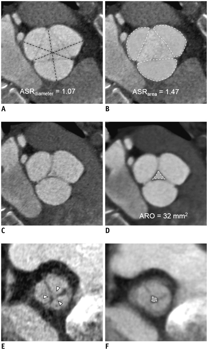
Fig. 3. 34-year-old female with type 1a AR.
A, B. Aortic cusp ASRdiameter (dashed lines) and ASRarea (dashed white lines) were measured on end-systolic phase. C, D. ARO was measured on end-diastolic phase. (C) Leaflets are not well demonstrable on 1-mm thickness image of AV in en-face view; therefore, (D) 5–10-mm-thick slices are used to measure ARO (dashed white line). E. On postoperative CT, four days after David operation, AV en-face view on end-diastolic phase demonstrated small central coaptation defect (arrowheads). F. Recurrent ARO was noted on thick-slice thickness (dotted-lined area). On same day, grade 3 AR was detected.

