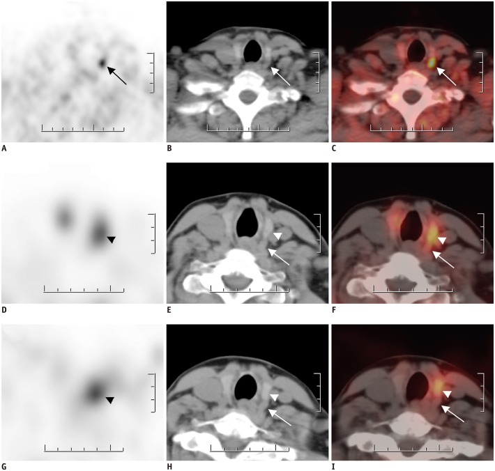
Fig. 3. FCH PET/CT (A–C) and MIBI SPECT/CT (D–I) findings for 48-year-old female with hyperparathyroidism (PTH level: 191 pg/mL).
FCH PET (A), CT (B), and fused PET/CT (C) images in FCH PET/CT reveal nodular lesion with intense FCH uptake (arrows) posterior to left thyroid lobe, which was confirmed to be parathyroid adenoma. Patient also showed papillary thyroid microcarcinoma in left thyroid lobe with no FCH uptake. In early MIBI SPECT (D), CT (E), and fused SPECT/CT (F) images, MIBI uptake was noted on left thyroid lobe. Moreover, MIBI uptake persisted in late SPECT (G), CT (H), and fused SPECT/CT (I) images (arrowheads). However, parathyroid lesion posterior to left thyroid lobe did not exhibit any MIBI uptake in early and late SPECT/CT scans (arrows). SPECT = single-photon emission computed tomography

