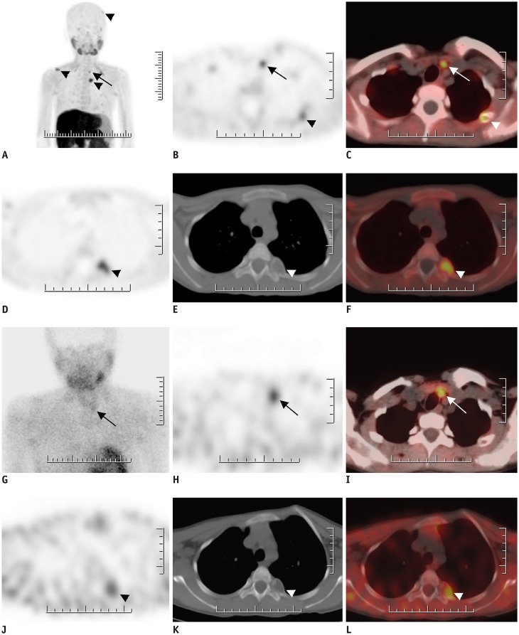
Fig. 5. 16-year-old female patient with chronic renal failure.
A–F. FCH PET/CT MIP image (A) reveals multiple foci of increased FCH uptake. Axial PET (B) and fused PET/CT (C) images on lower neck region reveal intense FCH uptake on left side that was found to be in accordance with parathyroid adenoma in surgical specimen (arrows). Brown tumors also demonstrate increased FCH uptake (arrowheads). G–L. MIBI SPECT/CT images of same patient. MIBI uptake at late planar (G), SPECT (H), and fused SPECT/CT (I) images reveal MIBI uptake on parathyroid adenoma (arrows). Brown tumors also demonstrate increased MIBI uptake (arrow heads).

