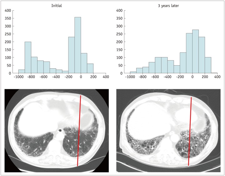
Fig. 4. Interval CT images of 70-year-old man with idiopathic pulmonary fibrosis.
Compared to histogram of initial CT scan, histogram of CT scan obtained three years later demonstrates right-side shifting of Hounsfield unit pixels due to microscopic interstitial fibrosis, suggesting progression of idiopathic pulmonary fibrosis.

