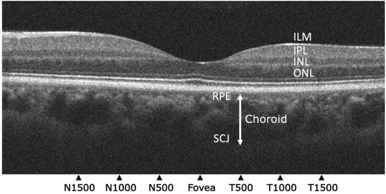
Figure 1.
Optical coherence tomography of the retina and choroid. Choroidal thickness was measured from the retinal pigment epithelium (RPE) to the sclerochoroidal junction (SCJ; white double-headed arrow). These locations were 1500 μm, 1000 μm, and 500 μm nasal and temporal to the fovea, and underneath the foveal center. Representative layers of the retina: ILM = Internal Limiting Membrane; IPL = Inner Plexiform Layer; INL = Inner Nuclear Layer; ONL = Outer Nuclear Layer (12).

