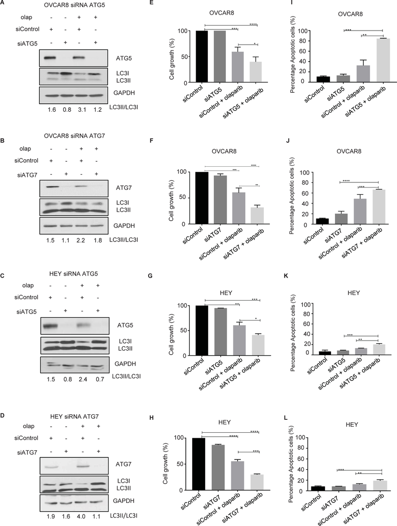
Figure 3. Inhibition of olaparib-induced autophagy by knocking down ATG5 and ATG7 decreases cell viability and enhances apoptosis.
OVCAR8 and HEY cells were transfected with ATG5 or ATG7 siRNA and treated with olaparib (5 μM). A-D. Knockdown efficacy was observed by western blot analysis. E-H. 4,000 cells/well were plated in 96-well plates, then sequentially transfected with ATG5 or ATG7 siRNA. After 24 hours cells were treated with olaparib (5 μM) for 5 days. Cells were fixed and stained by sulforhodamine B. I-L. Transfected cells were treated with olaparib for 96 hours. After incubation with Annexin V-FITC in a buffer containing propidium iodide, cells were analyzed using flow cytometry. Data represents results from at least two experiments.

