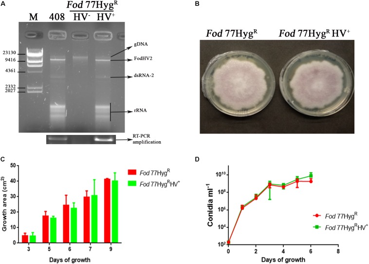
FIGURE 6.
Biological effect of FodHV2 on Fusarium oxysporum f. sp. dianthi. (A) Agarose gel electrophoresis of the dsRNA extracts from the strains Fod 408 (the originally infected with FodHV2), Fod 77HygRHV– (not infected), and Fod 77HygRHV+ (the new infected strain to which the virus was transferred), and RT-PCR products obtained using these dsRNA extracts and specific primers for the RdRp segment of FodHV2. M: molecular weight marker II (Roche Diagnostics). (B) Colony morphology of Fod 77HygR and Fod 77HygRHV+ after 9 days of growth on PDA. (C) Two dimensional colony growth rate of Fod 77HygR and Fod 77HygRHV+. The colony area was measured at 3, 5, 7, and 9 days of growth on PDA with hygromycin at 25°C in the dark. Values are the average area of five colonies. (D) Conidiation rate of Fod 77HygR and Fod 77HygRHV+ in liquid medium. Isolates were cultured in casein hydrolyzed medium, and conidia counted at 1, 2, 3, 4, 5, and 6 days of growth. Vertical lines in C and D represent the standard error.

