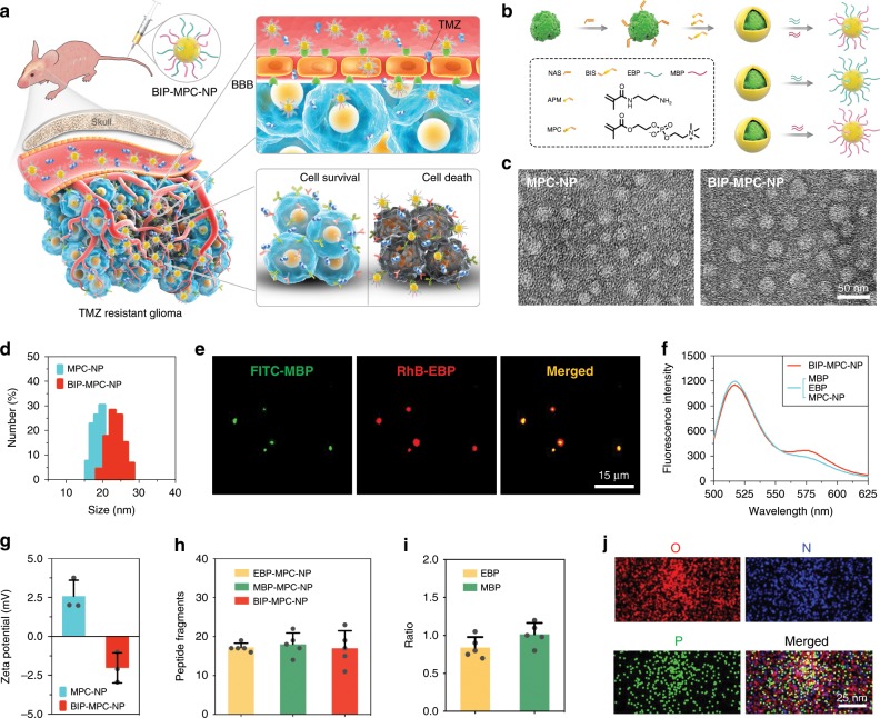
Fig. 1. The synthesis and characteristics of nanoinhibitors.
a Schematic illustration and delivery process of the nanoinhibitor, BIP-MPC-NP, enhancing TMZ sensitivity against glioma via intravenous injection. b The schematic diagram revealing the establishment and structure of nanoinhibitors. c The transmission electron microscope (TEM) images of the nanoinhibitors. Scale bar = 50 nm. d The dynamic light scattering (DLS) measurements of BIP-NP and BIP-MPC-NP. e Fluorescence images presenting the co-localization of FITC-MBP and RhB-EBP for each BIP-MPC-nanoparticle. Scale bar = 15 μm. f Förster resonance energy transfer (FRET) analysis of BIP-MPC-NP and MBP/EBP/MPC-NP. g The zeta potential data of BIP-NP and BIP-MPC-NP (n = 3). h The quantitative analysis of peptide fragments per nanoinhibitor (n = 5). i The ratio of EBP and MBP on the nanoparticles (n = 5). j TEM micrograph exhibited the oxygen element (O), nitrogen element (N) and phosphorus element (P). Scale bar = 25 nm. The error bars in g, h and i represent the S.D. of three or five measurements.

