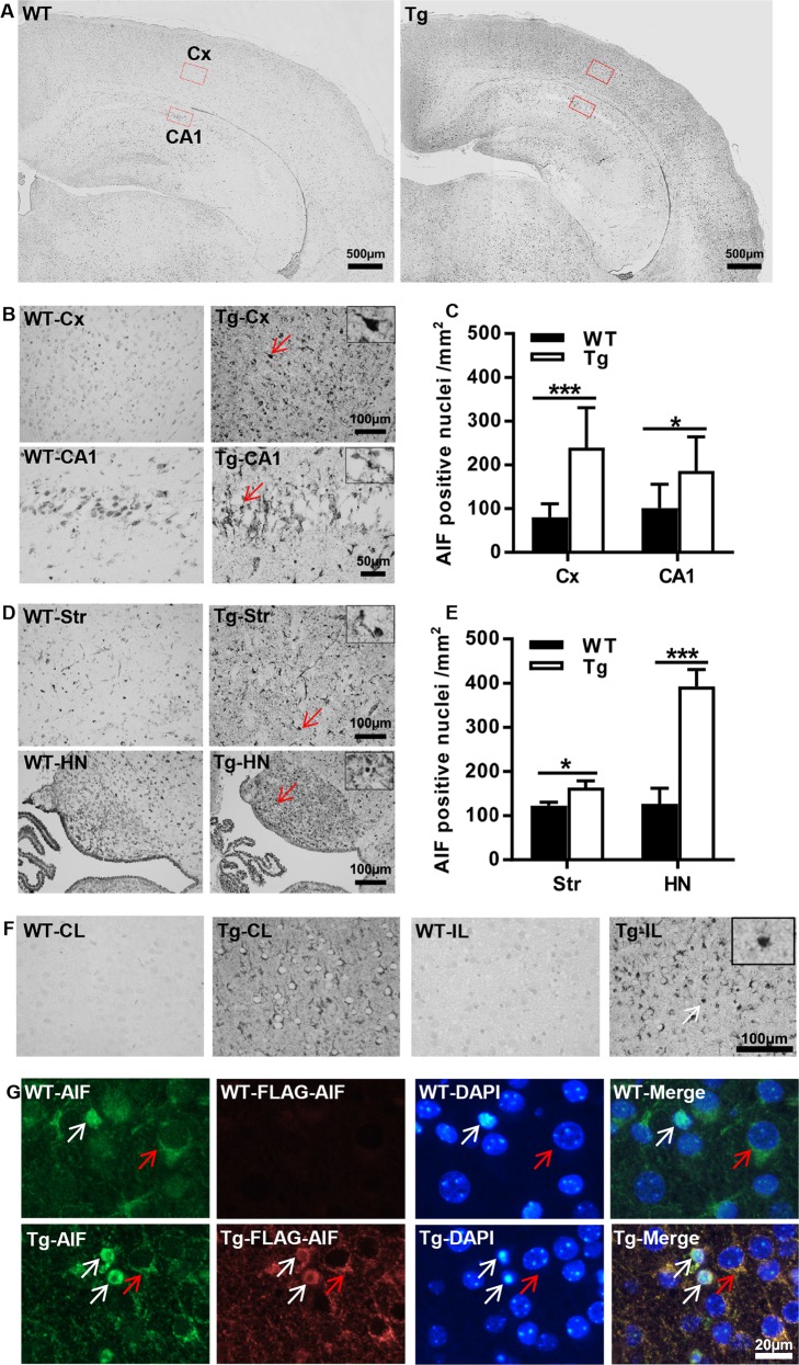
Fig. 4. AIF overexpression increased AIF nuclear translocation in the brain after HI.
a Representative panoramic images of AIF staining showing the cortex and hippocampus in WT and AIF Tg mice at 24 h post-HI. b Higher magnifications of AIF staining images show AIF-positive nuclei in the cortex (Cx) area (upper panels) and hippocampal cornus ammonis 1 (CA1) area (lower panels) in WT and AIF Tg mice at 24 h post-HI. c Quantification of AIF-positive nuclei in WT and AIF Tg mice at 24 h after HI in the cortex (80.4 ± 30.7 cells/mm2 vs. 239.2 ± 91.8 cells/mm2, 95% confidence interval (CI) 78.5–239.1, respectively, n = 8/group, ***p < 0.001) and CA1 (101.1 ± 54.6 cells/mm2 vs. 186.1 ± 78.4 cells/mm2, 95% CI 4.6–165.3, respectively, n = 8/group, *p < 0.05). d Higher magnifications of AIF staining images showing the AIF-positive nuclei in the striatum (Str) (upper panels) and habenular nuclei (HN) (lower panels) in WT and AIF Tg mice at 24 h post-HI. e Quantification of AIF-positive nuclei in in WT and AIF Tg mice at 24 h after HI in the striatum (122.8 ± 21.8 cells/mm2 vs. 163.7 ± 42.6 cells/mm2, 95% CI 49.9–131.8, respectively, n = 8/group, *p < 0.05) and habenular nuclei (126.6 ± 100.0 cells/mm2 vs. 392.7 ± 106.6 cells/mm2, 95% CI 175.3–357.0, respectively, n = 8/group, ***p < 0.001). f FLAG staining in the cortex showing the expression of exogenous AIF in Tg mice, but not in WT mice (left panels, WT-CL and Tg-CL), and the nuclear translocation of exogenous AIF (right panels, WT-IL and Tg-IL). g Immunofluorescence staining of AIF (green) and FLAG (red) showing the nuclear translocation of both endogenous AIF and exogenous AIF at 24 h after HI (white arrows point to the injured cells, red arrows point to the normal cells).

