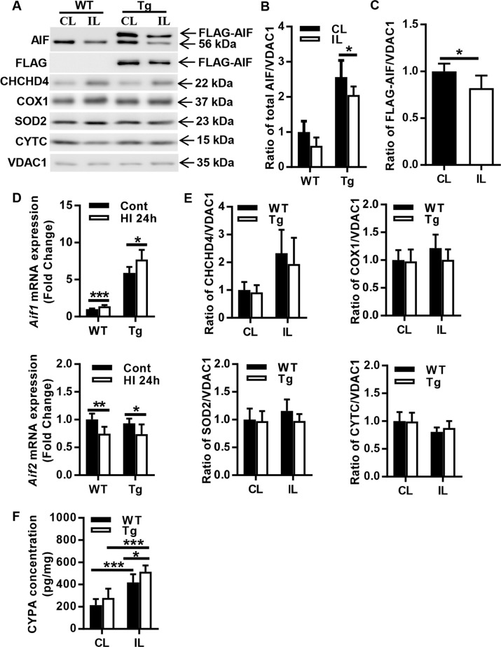
Fig. 6. Changes in mitochondria-related proteins in the cortex after HI.
a Representative immunoblotting of AIF, FLAG, CHCHD4, COX1, SOD2, and CYTC in the mitochondrial fraction of cortical tissue from the CL and IL hemispheres of WT and AIF Tg mice at 24 h after HI. b Quantification of AIF protein in the mitochondrial fraction from the CL and IL hemispheres of WT and AIF Tg mice at 24 h after HI (n = 6/group). A greater reduction in AIF from the mitochondria was seen in the IL of AIF Tg mice. c Quantification of FLAG-AIF protein in the mitochondrial fraction from the CL and IL hemispheres of AIF Tg mice at 24 h after HI (n = 6/group). FLAG-AIF was significantly released from the mitochondria in the IL of AIF Tg mice. d The mRNA expressions of Aif1 and Aif2 were determined by RT-qPCR in cortical tissue of WT and AIF Tg mice at 24 h post-HI. Aif1 expression was increased in both WT and AIF Tg mice, but Aif2 expression was decreased in both WT and AIF Tg mice (n = 6/group). e Quantification of mitochondria-related proteins (CHCHD4, COX1, SOD2, and CYTC) did not show any significant differences between WT and AIF Tg at 24 h after HI, but there was increased CHCHD4 expression in the IL of both WT and AIF Tg mice (n = 6/group). f CYPA in the nuclear fraction of cortical tissue was significantly increased in the IL hemisphere of both WT and AIF Tg mice at 24 h after HI. The increase was more obvious in AIF Tg mice (n = 6/group). *p < 0.05, **p < 0.01, ***p < 0.001.

