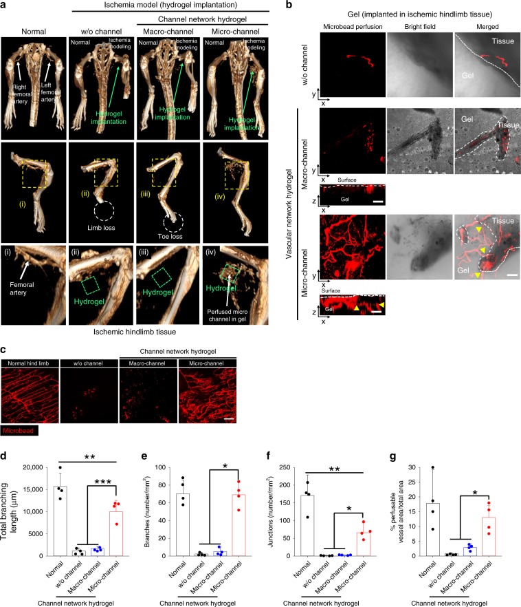
Fig. 2. Promotion of host vessel ingrowth and perfusion connection with channel networks.
a MicroCT images of arterial vasculature in ischemic tissue of mouse hindlimb at day 14 post hydrogel implantation (green dot box). i–iv Middle and bottom rows: high magnification images of the hydrogel implantation sites. Confocal images of harvested b whole channel network hydrogel and c hindlimb tissues post-perfusion of red FluoroSpheres, through the left ventricle at day 14 indicate ingrowth (yellow arrows) of host blood vessels and perfusion connection with the microchannel network (red). Scale bars = 100 μm. Quantification of microvasculature structural parameters: d total branching length, e branch number, f junction number, and g perfusable vessel area per field of view (FOV) at ischemic hindlimb tissue (N = 4). Dots represent each animal. Data presented are mean ± SEM. Statistical significances are determined using one-way ANOVA with Tukey post-hoc pairwise comparisons; *p < 0.05, **p < 0.01, and ***p < 0.005 between lined groups. Source data are provided as a Source Data file.

