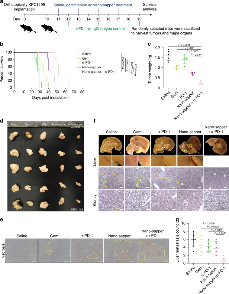
Fig. 7. Nano-sapper synergized with α-PD-1 to suppress tumor growth and metastasis in KPC1199 PDAC model.
a Six-week-old male C57BL/6 mice were orthotopically inoculated with KPC1199 cells (1 × 10⁶) on day 0. Saline, Gem (15 mg/kg) and Nano-sapper (MP = 13.9 mg/kg, 25 μg plasmid per mouse) were i.v. administered every other day and α-PD-1 or IgG isotype (200 μg per mouse) were i.p. administered every 2 days (n = 12 mice). One day after the final α-PD-1 treatment, five mice were randomly sacrificed to extract tumors and major organs, while the rest mice were continually subjected to survival analysis. b Mice survival curves in each treatment group (n = 6 mice). c, d Tumor inhibition of different treatments (n = 5 mice). e H&E staining histology of tumor necrosis. Scale bars, 100 μm. f, g H&E staining histology of tumor metastasis in liver and kidney. Nodules of metastasis were indicated by white arrows and focal necrosis were indicated by yellow arrows (n = 5 mice). Data are presented as mean ± s.d. One-way ANOVA with Bonferroni multiple comparisons post-test was used for (c) and (g). Kaplan–Meier analysis with log-rank Mantel-Cox test (two-sided) was used for (b). Error bars represent s.d.

