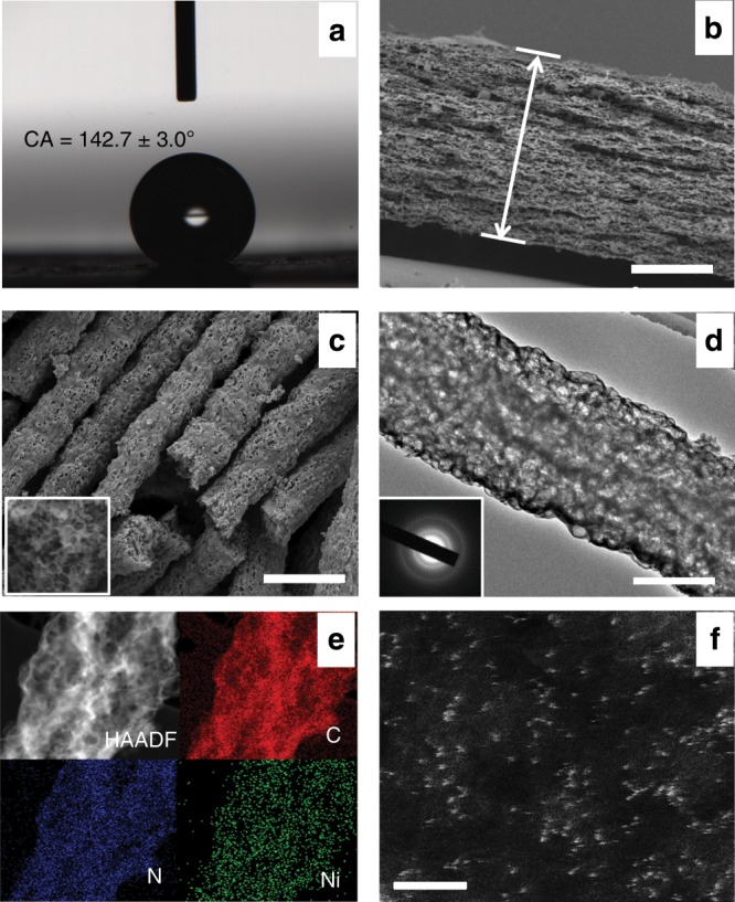
Fig. 2. Morphological and structural characterizations of NiSA/PCFM.

a The water contact angles of NiSA/PCFM spraying with a little amount of Nafion solution. b Cross-sectional and c high-resolution SEM images of NiSA/PCFM. d HR-TEM image of NiSA/PCFM, the inset of (d) displays the lattice fringe. e EDS mapping of an independent NiSA/PCFM nanofiber. f Magnified HAADF-STEM images of NiSA/PCFM, those white dots are supposed to be Ni single atoms. Scale bars, 100 μm (b), 1 μm (c), 0.5 μm (d), and 2 nm (f).
