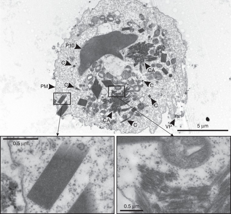
Fig. 2. TEM studies of TbIMPDH crystals.
Transmission electron micrograph of a Sf9 cell 6 days p.i. showing multiple TbIMPDH crystals with varying dimensions. Sub-micron crystals are indicated by “C”. Signs of infection are clearly visible (baculoviral P10 protein - “P10”, viral particles - “VP”). TbIMPDH not only crystallizes in needle-shaped crystals characterized by a regular crystal lattice (left inset), but also seems to create irregular crystalline assemblies (“CA”, right inset) that display fragmented crystal lattices and spread over several µm within the cytoplasm. PM plasma membrane.

