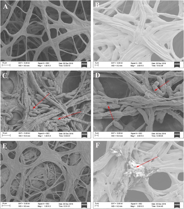
Fig. 7.
SEM micrographs of antagonistic bacteria interacting with hyphae of Fof on PDA medium. a, c, e Magnification: 1000×. b, d, f Magnification: 2000×. a, b Fof hyphae alone; c, d Fof hyphae treated with strain X-1 (red arrows show Fof hyphae surrounded by X-1); e, f Fof hyphae treated with strain Z-1 (red arrows indicate Fof hyphae damaged by Z-1)

