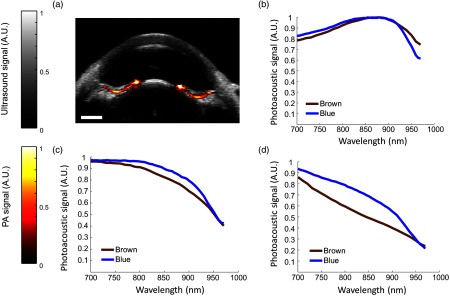
Fig. 5.
Comparison of measured PA spectra in blue and brown eyes. (a) Overlay of ultrasound (gray) and phototacoustic (red) images in a blue porcine eye. PA signal was localized to the posterior iris. (b–d) Spectra were separated based on results from brown porcine eyes. (b) A modified melanin spectrum, resembling that of oxygenated hemoglobin, was isolated to the posterior iris in both eye colors and was the dominant spectrum observed in blue eyes. (c and d) Other types of spectra were observed in blue eyes, but were not localized to a consistent location. (d) The PA spectrum resembling melanin (b) was most prevalent in brown eyes. .

