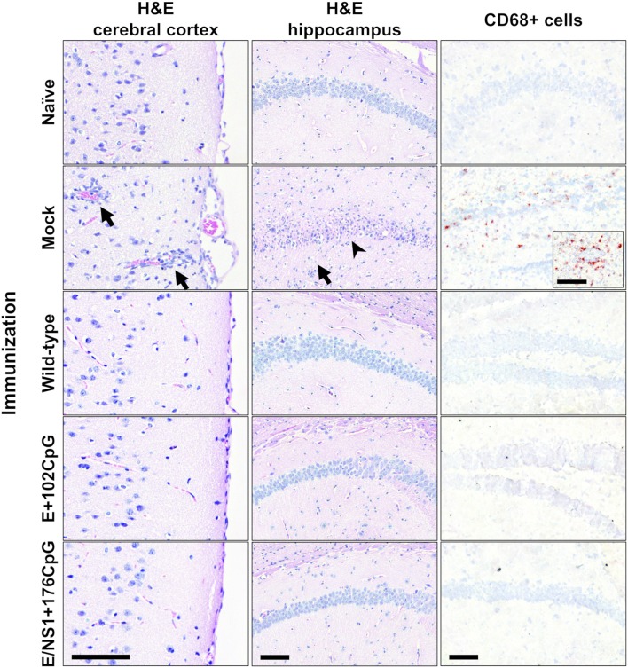
Figure 11.
Histopathology and immunohistochemistry in mouse brains. H&E: Cortical monocyte infiltration, inflamed cortical blood vessels (arrows), hippocampal monocyte invasion (arrow), and necrosis of hippocampal neurons (arrowhead) were detected only in mock-immunized mice. Immunohistochemistry: CD68-positive cells were detected in brains from only mock-immunized mice. Brains from all control and experimental animals (Figure 10) were tested. Scale bars are 100 μm.

