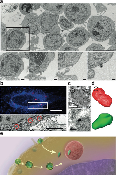
Figure 3 |.

Imaging the large and small by FIB-SEM. (a) Very large fields of view including many HIV-infected cells (top) can be imaged at resolutions high enough to visualize individual virions in the same image (bottom; panels show increasing magnifications of the boxed region above)55. (b) Using fiducial markers, confocal (top) and FIB-SEM image volumes (bottom, corresponding to the boxed area above) can be registered; correlative fiducials are denoted by red channels (top) and areas circled in red (bottom). (c,d) FIB-SEM image (c) and segmentation (d) of individual HIV cores (dark signal in c (top and bottom), segmented green region in d) associated with TRIM5-α bodies (fuzzy gray body in c (top and bottom), segmented red region in d) in the cytoplasm of infected cells. With CLEM one can locate these cores and visualize them in three dimensions, achieving imaging across a volume scale of 109 in a single FIB-SEM imaging experiment60. (e) A model for HIV transport to the nucleus; the postfusion intermediate of the viral core is stabilized by association with the surface of TRIM5-α bodies. Scale bars, 9 μm (a, top); 1.2 μm, 600 nm, 300 nm and 150 nm (a, bottom, from left to right); 10 μm (b, top); 5 μm (b, bottom); 400 nm (c, top); and 100 nm (c, bottom). Panel a copyright © American Society for Microbiology (Journal of Virology, Vol. 88, 2014, 10327–10339, doi:10.1128/JVI.00788-14). Panels b–d reprinted from Journal of Structural Biology, Vol. 185, Narayan, K. et al., “Multi-resolution correlative focused ion beam scanning electron microscopy; applications to cell biology,” 278–284, Copyright 2014, with permission from Elsevier.
