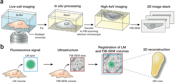
Figure 4 |.

3D imaging of specific targets with correlative LM and FIB-SEM. (a) LM of a biological sample grown on or attached to an alphanumerically coded gridded coverslip produces a ‘coordinate map’ whose fidelity is maintained after resin embedding in situ, allowing location of the ROI for FIB-SEM imaging, (b) Fluorescence microscopy of tagged targets in the biological sample yields a 2D or 3D ‘target map’. FIB-SEM images subsequently acquired from the same sample can then be registered to the fluorescence image, allowing the user to reliably obtain the nanoscale 3D structures of specific fluorescent targets in a sample.
