
Figure 5 |.
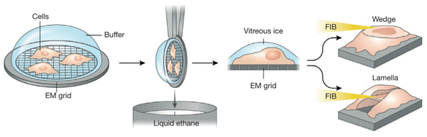
FIB thinning and lift-out of specimens for cryo-imaging. Cells grown or pipetted on TEM grids are plunge-frozen in liquid ethane and transferred to an FIB-SEM under cryogenic temperatures. A chosen area is FIB-milled tangentially either from one direction to produce a wedge or from above and below to produce a lamella, revealing the region to be imaged. This region, still encased in vitreous ice, is now thin enough to be imaged in the FIB-SEM in scanning transmission electron microscopy mode, or it can be transferred to a transmission electron microscope under cryogenic conditions and imaged at high resolution.