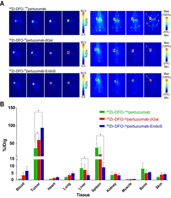
Figure 4.
(A) Planar (left) and maximum intensity projection (MIP, right) PET images of huNSG mice bearing subcutaneous BT474 xenografts collected between 24 and 144 h after the administration of the three radioimmunoconjugates (209 - 218 μCi, 7.7 - 8.1 MBq, 80 - 83 μg, in 200 μl 0.9% sterile saline); (B) Biodistribution data for 89Zr-DFO-nsspertuzumab, 89Zr-DFO-sspertuzumab-βGal, and 89Zr-DFO-sspertuzumab-EndoS 144 hours following administration in huNSG mice bearing subcutaneous HER2-expressing BT474 xenografts. T = tumor; L = liver; S = spleen; B = bone. * = p < 0.05.

