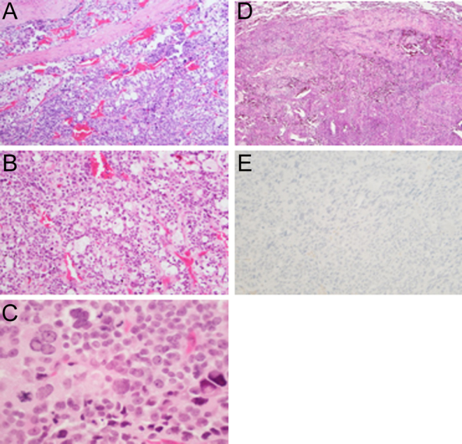
Figure 4.

(A) Low power of the adrenocortical carcinoma involving the fibrous capsule (150× magnification, haemotoxylin and eosin staining). (B) Higher magnification of the tumor shows small cells with dark nucleus and scant cytoplasm intermixed with lipid-rich cells with prominent clear cytoplasm (200× magnification, haemotoxylin and eosin staining). (C) The tumor cells show variation in nuclear size and nuclear pleomorphism. Mitotic figures were present (250× magnification, haemotoxylin and eosin staining). (D) Metastatic tumor in the lung. The tumor showed the same characteristics of the primary adrenal lesion (150× magnification, haemotoxylin and eosin staining). (E) Immunohistochemical staining of the tumor was negative for chromogranin (150× magnification).

 This work is licensed under a