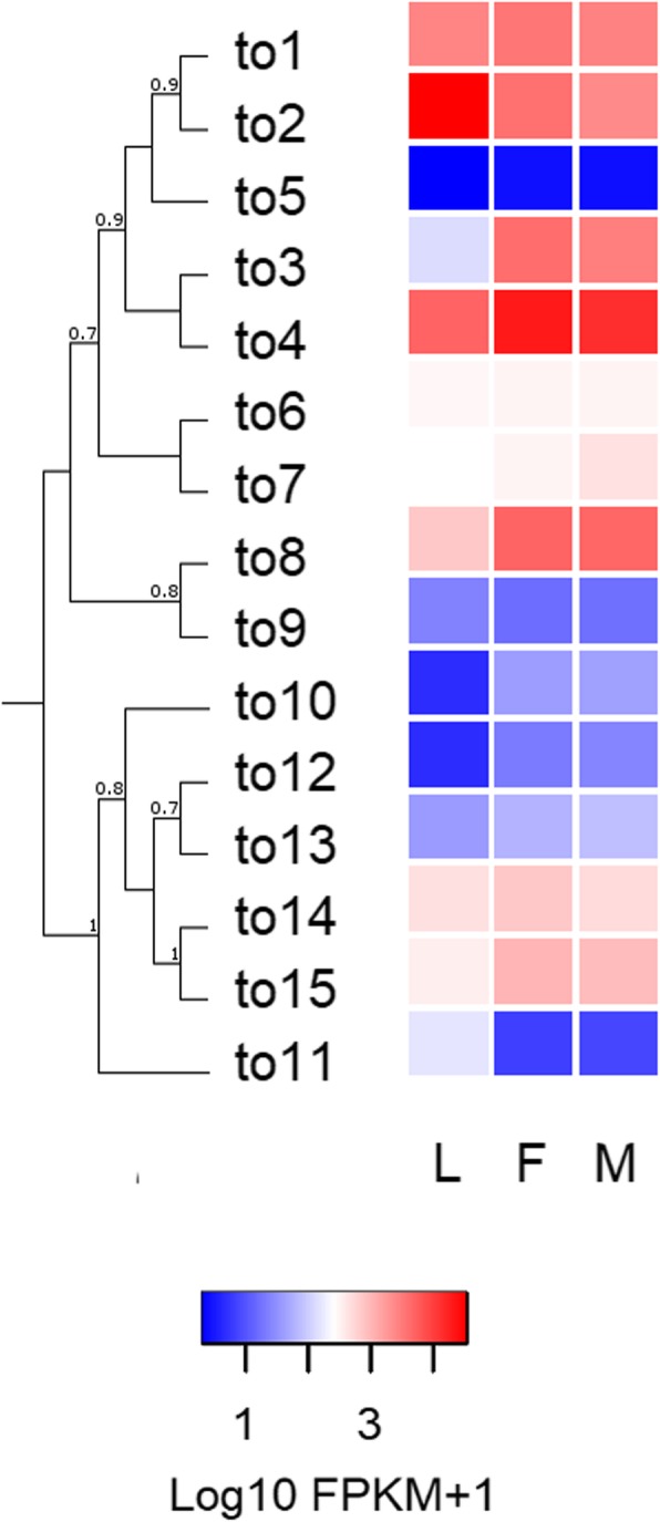
Fig. 4.

Heat map comparing the expression levels of R. prolixus to genes in the antennae of larvae (L), female (F) and male (M) adults. Expression levels (displayed as Log10 FPKM + 1) represented by means of a colour scale, in which blue/red represent lowest/highest expression. The evolutionary history of R. prolixus takeout genes was inferred by using the maximum likelihood method in PhyML v3.0. The support values on the bipartitions correspond to SH-like P values, which were calculated by means of the aLRT SH-like test. The LG substitution amino-acid model was used
