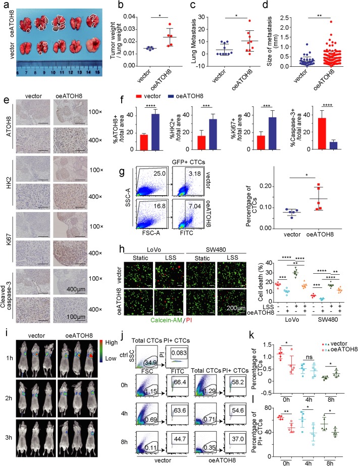
Fig. 2.
ATOH8-overexpressing colorectal tumour cells tend to survive and metastasize in the circulation. a Stably transfected SW480 cells with GFP labelling were injected intravenously into nude mice and then lung metastasis model was established 4 weeks later. The gross view of lung metastasis from nude mice in vector or ATOH8-overexpressing groups was presented. b The statistical result of the weight rate of lung metastasis/ lung tissue in vector or ATOH8-overexpressing groups. c, d The statistical result of metastatic nodule numbers (c) and sizes (d) in the lungs from vector or ATOH8-overexpressing groups. e, f Immunohistochemistry (e) and quantification (f) graph of the ratio of ATOH8+, HK2+, Ki67+ and cleaved caspase 3+ cells of tumour samples from the ATOH8 overexpression group and control groups. g Left, the GFP (+) SW480 cancer cells percentage in the blood of lung metastatic nude mice was analysed by flow cytometry. Right, the statistical result of the percentage of GFP (+) SW480 was presented. h Live/dead cell vitality assay of suspended LoVo and SW480 cells were treated with LSS (10 dyn/cm2, 30 min). Representative fluorescence images (Left) and quantification of dead cells (Right) were displayed. Red in the images denotes dead cells, while green denotes live cells. i Vector or ATOH8-overexpressing SW480 cells with luciferase were injected intravenously, and in vivo imaging was performed at 1, 2 and 3 h after the injection. j Vector or ATOH8-overexpressing SW480 cells with GFP labelling were injected intravenously, and flow cytometric cell apoptosis assays was performed at 0, 4 and 8 h after the injection. Different groups of representative flow cytometry diagrams were displayed. k, l The statistical result of the number of total CTCs (k) and apoptotic CTCs (PI+ CTCs, l) based on j. *P < 0.05, **P < 0.01, ***P < 0.001 and ****P < 0.0001

