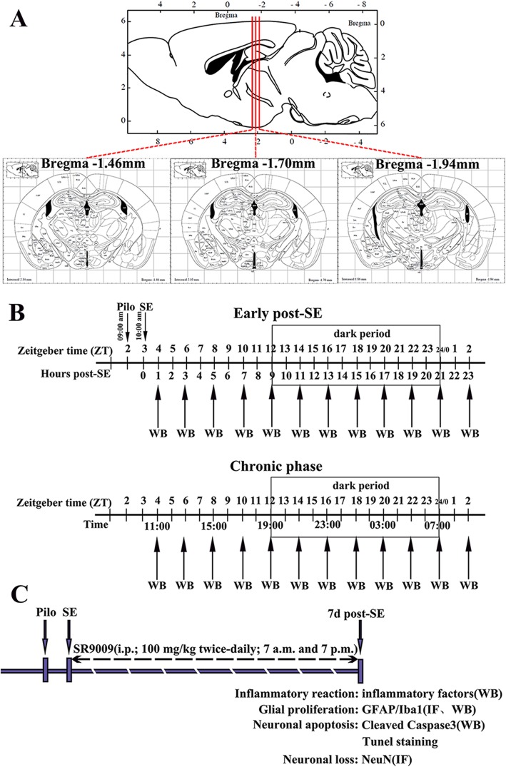
Fig. 1.
Schematic diagram of the experimental design. a Three coordinate positions of coronal sections of mouse brain. b Pilocarpine was injected at 9 a.m. (ZT 2). Mice that presented SE until 10 a.m. (ZT 3) were included in the experiments. The expression profiles of Rev-Erbα in the hippocampus and temporal neocortex of mice in the control group, early post-SE group, and chronic epilepsy group were determined by WB. Mice were euthanized every 2 h throughout the 24-h light-dark cycle at ZT 4, 6, 8, 10, 12, 14, 16, 18, 20, 22, 0, and 2 (5 controls and 5 experimental animals at each time point). The box indicates the dark period. c Schematic of SR9009 intervention therapy. SR9009 (100 mg/kg) was administered intraperitoneally twice daily (7 a.m. and 7 p.m.) for 7 consecutive days after SE. Then, we explored the effects of SR9009 on the inflammatory reaction, glial proliferation, neuronal apoptosis, and neuronal loss in the hippocampus 7 days after SE. PILO, pilocarpine; SE, status epilepticus; WB, western blotting; IF, immunofluorescence staining

