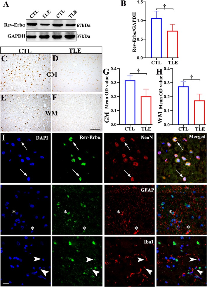
Fig. 2.
The expression patterns of Rev-Erbα in the temporal neocortex of patients with TLE and controls (CTLs). a The Rev-Erbα protein level was detected by western blotting in the temporal neocortex of TLE patients (n = 22) and controls (n = 10). b Densitometric analysis indicated that protein immunoreactivity for Rev-Erbα was decreased in TLE patients. In controls (n = 10), Rev-Erbα displayed moderate to strong IR in neurons within the gray matter (GM; c) and in glial cells within the white matter (WM; e). Rev-Erbα showed weak IR in corresponding regions in the TLE group (n = 22; d, f). The mean OD of Rev-Erbα in the GM (g) and WM (h) was significantly reduced in the TLE group compared with the control group. i Double-labeled immunofluorescence staining showed that Rev-Erbα was coexpressed with NeuN in neurons (arrows). Rev-Erbα was colocalized with GFAP in astrocytes (asterisks); Rev-Erbα and Iba1 were also colocalized (arrowheads). The data are expressed as the mean ± SD. †p < 0.001. Scale bar = 50 μm for c–f, i

