Fig. 1.
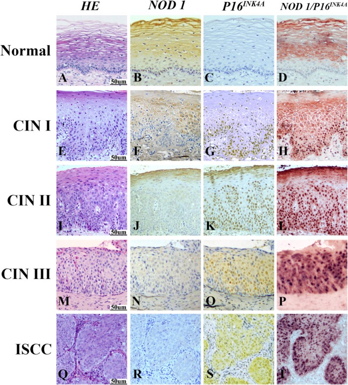
Expression of NOD1 and P16INK4A in normal cervix, CIN I, CIN II, CIN III and ISCC tissues detected by immunohistochemistry. Hematoxylin and eosin (HE) staining showed that in comparison to normal cervical squamous epithelium (a), atypical hyperplasia of squamous epithelium was restricted within lower third in CIN I (e), lower two-thirds in CIN II (i), or exceeded over lower two-thirds in CIN III (m), respectively. Carcinoma invaded into muscle tissue in ISCC (q). NOD1 showed the strongest immunoreactivity in normal cervical squamous epithelium (b), and the staining intensity of NOD1 gradually decreased from CIN I (f), CIN II (j) to CIN III (n). No NOD1 immunostaining was observed in cells within carcinoma nests (r). In contrast to NOD1, no p16INK4A staining was detected in the normal cervical tissue (c). The expression of p16INK4A gradually increased from CIN I (g), CIN II (K) to CIN III (o), and p16INK4A positive cells were diffusely distributed within the atypical hyperplasia tissue. Diffuse and strong staining of p16INK4A was observed in tumor epithelial cells of carcinoma nests in ISCC (S). NOD1 staining was only detected in normal cervical squamous epithelium (d) with double staining of NOD1 (red) and p16INK4A (black), and NOD1 staining was not observed within atypical hyperplasia cells of CINs (H, L, P), where p16INK4A was positive. Strong p16INK4A staining but no NOD1 staining was observed in the tumor cells of carcinoma nests (t)

