Fig. 3.
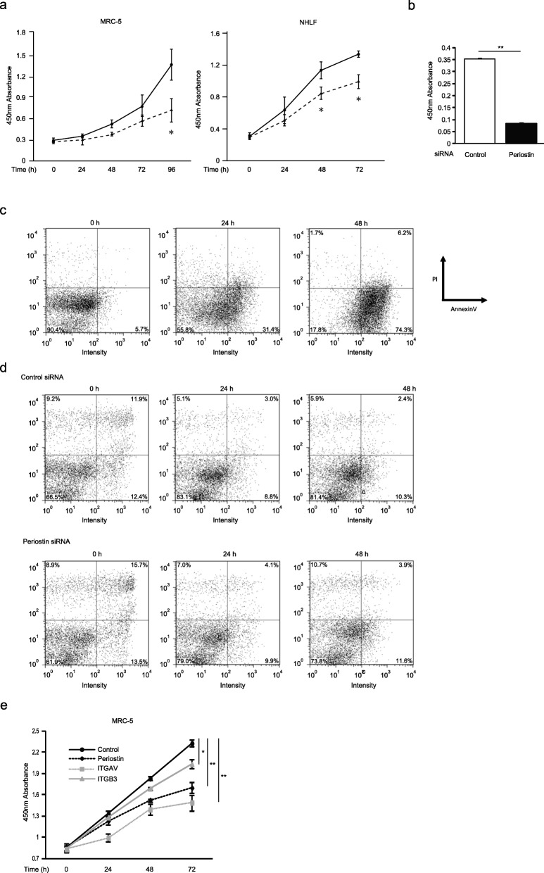
Periostin silencing slows cell proliferation in lung fibroblasts. a, b, e The growth curves of MRC-5 cells or NHLF. a The cells with (dashed line) or without (solid line) treatment of periostin knockdown were plated at a density of 1.0 × 10⁴ cells/well in 96-well plates. b The cells were treated with control siRNA or periostin siRNA for 48 h and pulsed with BrdU. After 12 h, the incorporation of BrdU was counted. Values are mean ± SD of three independent experiments. e The cells treated with siRNA for control (black solid line), periostin (black dashed line), αV integrin (gray dashed line), or β3 integrin (gray solid line) were plated at a density of 1.0 × 10⁴ cells/well in 96-well plates. The cell numbers were evaluated at the indicated times. Values are mean ± SD of three independent experiments. *P < 0.05, **P < 0.01. c, d Flow cytometric analysis of annexin V (horizontal) and PI (vertical) labeling is depicted. The proportions of each fraction have been inserted. The same experiments were performed twice. MRC-5 cells were treated with 50 μg/mL cycloheximide and 50 ng/mL TNF-α (c) or siRNA for periostin (d) for the indicated times

