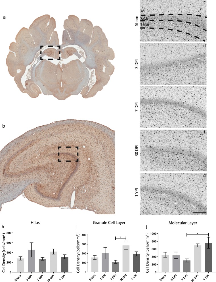
Fig. 5.
Microglia density increases in the anterior hippocampus over time following mild TBI. Microglia, stained with Iba-1, in pig coronal sections containing anterior hippocampus (a) with an enlarged call out box (b). The molecular layer (ML), granule cell layer (GCL), and Hilar region of the hippocampus were identified for each specimen and a representative image of Iba-1 staining for each experimental group is displayed (c–g) (scale = 200 μm). Microglia cell counts are quantified and graphed (h–j). Overall, biphasic trends can be observed as microglia tended to increase at 3 days post-injury (DPI), then decrease at 7 DPI, and finally increase again at chronic timepoints. i There is a significant increase in microglia from 7 DPI to 30 DPI in the GCL (p = 0.0312) and j a significant increase in microglia from 7 DPI to 1 year post-injury (YPI) in the ML (p = 0.0348)

