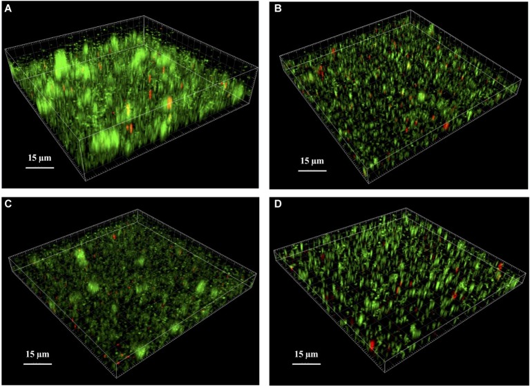
Figure 4.
Analysis of S. lugdunensis biofilm by confocal laser scanning microscopy (CLSM). The 24 h mature biofilms of (A) S. lugdunensis WT, (B) ΔlytSR, (C) ΔlytSR (pCU1:lytSR), and (D) ΔlytSR (pCU1) were visualized after Live/Dead staining under CLSM. Live cells stained with SYTO®9 appear in green, while dead cells stained with propidium iodide are in red. Three-dimensional structural images were reconstructed, and the amount of fluorescence of viable and dead cells was determined using Imaris software. The figures represent one of three independent experiments.

